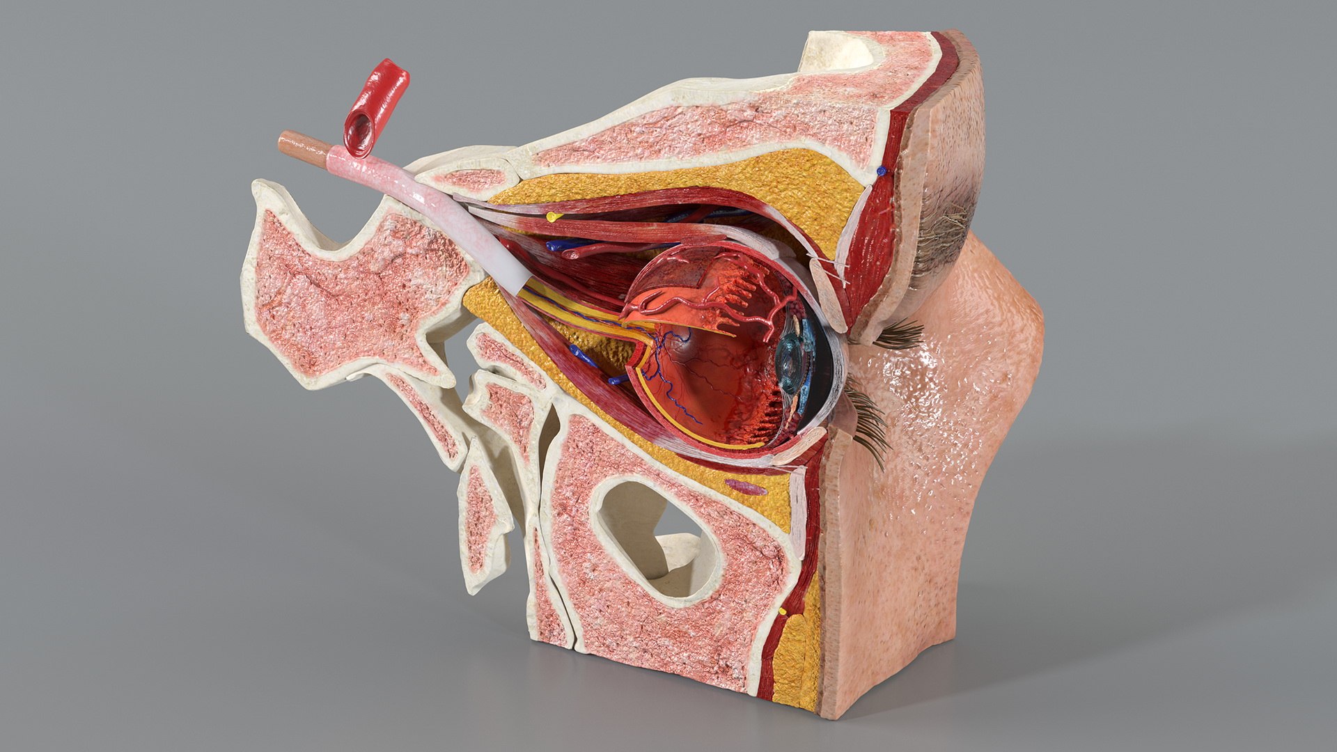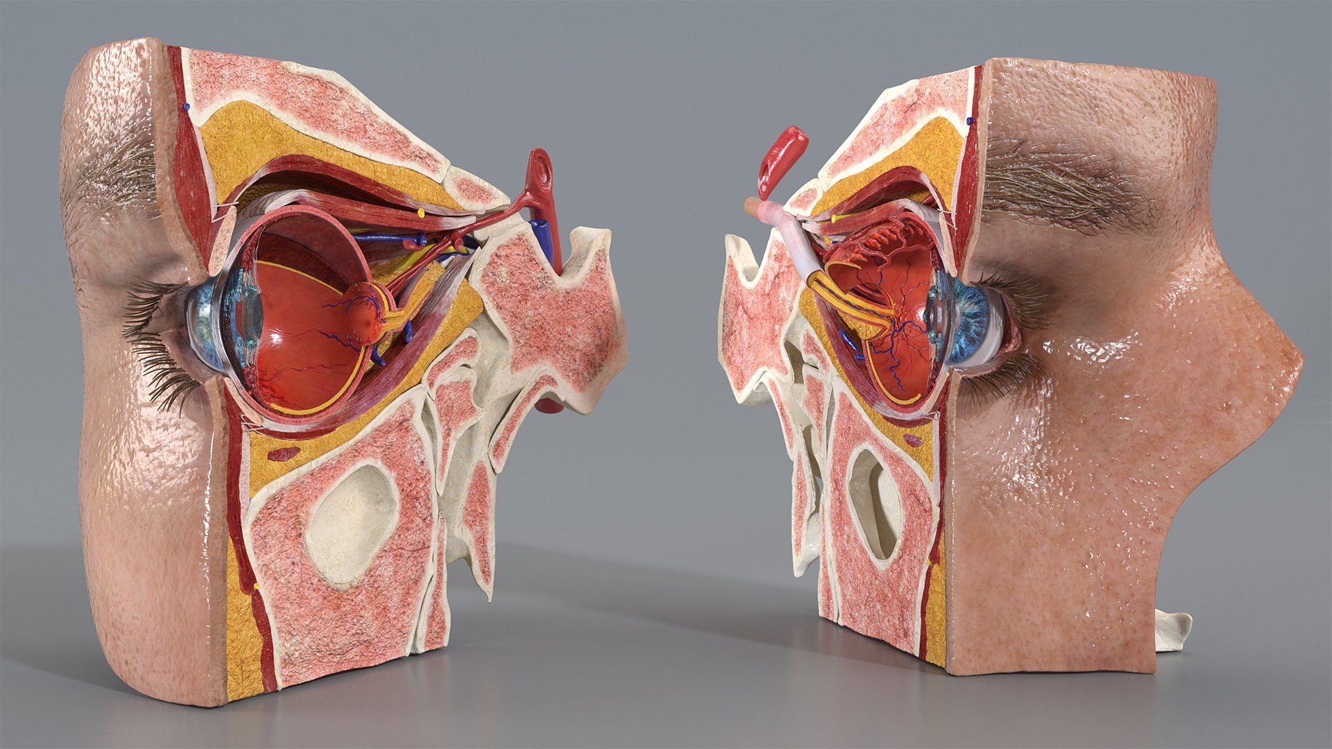
A sectioned eye is how the organ gets taught, because almost everything important is inside: the lens behind the iris, the vitreous filling the globe, the retina lining the back and the optic nerve leaving it. A whole eye shows a sphere; a cut one shows an optical instrument.
The mesh runs 212,864 polygons and 214,566 vertices in quads and tris with non-overlapping UVs, and ships with 45 texture maps - 40 at 4096x4096 and 5 at 2048x2048, which is what the retinal vasculature and the iris detail demand.
At $249 and built to StemCell specification, it suits ophthalmology and optometry education, medical and patient communication, courseware and textbook illustration, device marketing and broadcast medical work. On sale since 2020.
Questions About This Model
Can the Eye Anatomy Cross-Section model be modified after purchase?
Yes. The Royalty Free license allows editing the geometry, retopologising, changing materials and adapting the asset to a project. What it does not allow is reselling or redistributing the model file itself.
Where can I buy the Eye Anatomy Cross-Section 3D model?
It is sold through TurboSquid, where this listing has been available since 2020. Purchase, download and licensing are handled by TurboSquid; delivery is immediate after checkout.
What texture resolution ships with the Eye Anatomy Cross-Section?
45 maps at 4096 x 4096 and 2048 x 2048, with yes, non-overlapping UVs. That is enough for close framing without resampling.
What is the Eye Anatomy Cross-Section model typically used for?
The listing names medical visualization, medical simulation and software demos as the main applications. In practice it also works anywhere a finished medical object is needed without modeling it from scratch.