Skeleton & Bone 3D Models
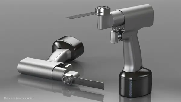
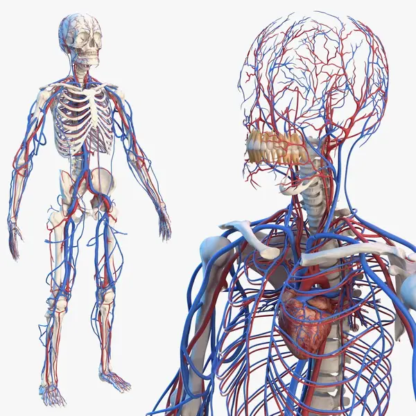
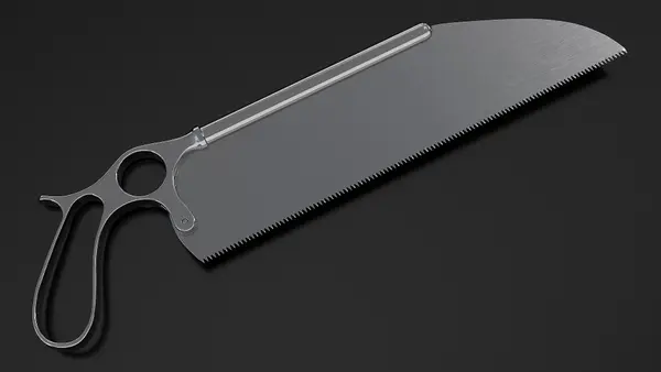
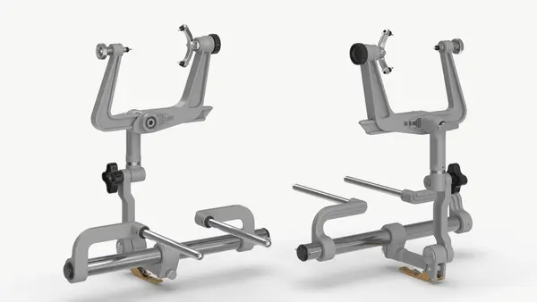
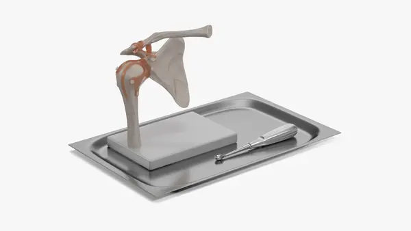
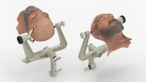
Skeletons and bones for anatomy teaching, orthopedics, forensic and story scenes: complete male and female skeletons, torsos, ribcages, pelvises, arms, legs, hands, feet and knees, and a skull that comes apart. Some figures add organs, vessels and nerves to the bones. Surgical pieces sit alongside, such as skull clamps, a bone saw and a cranial mesh implant.


About Skeleton & Bone 3D Models
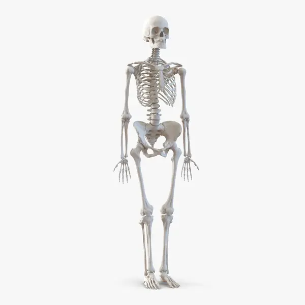
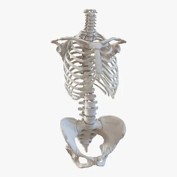
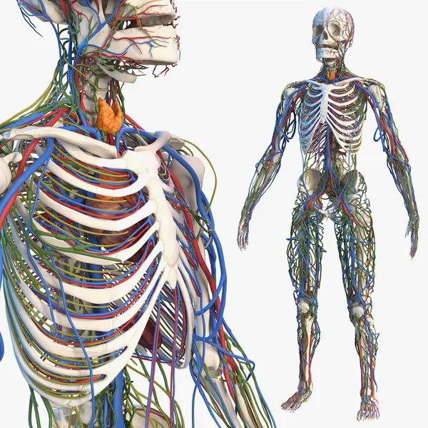
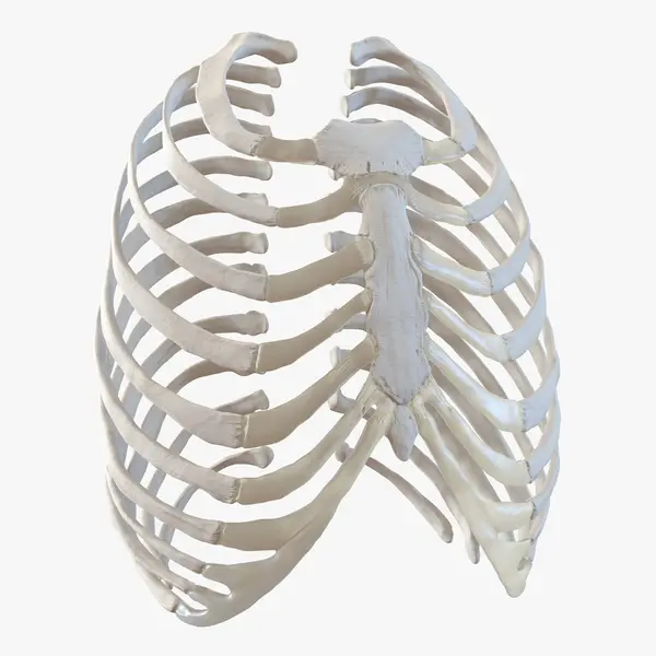
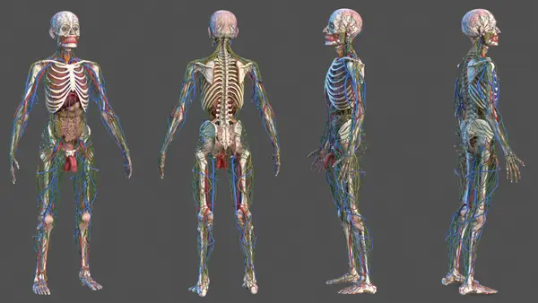
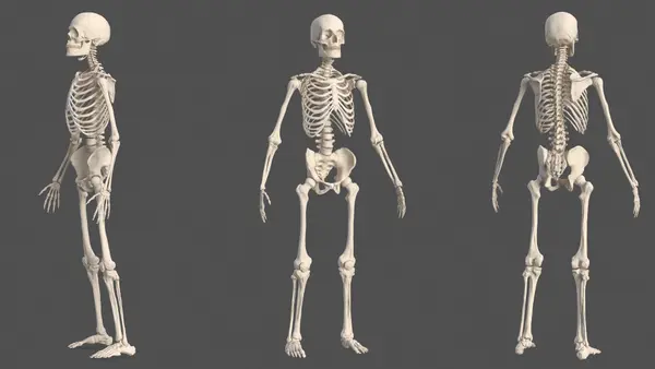
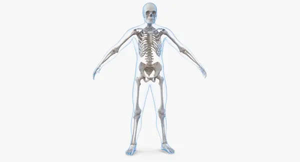
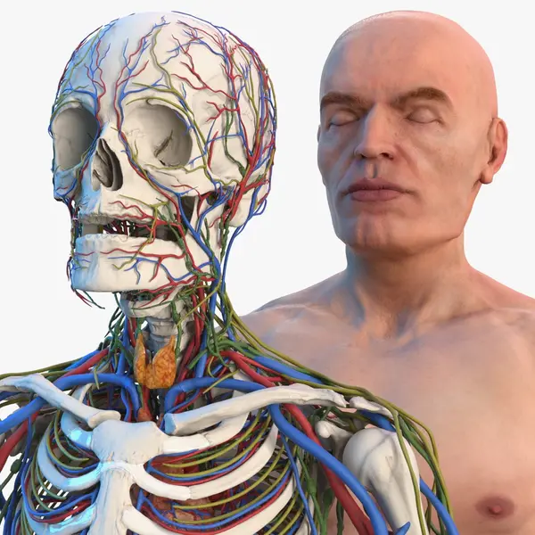
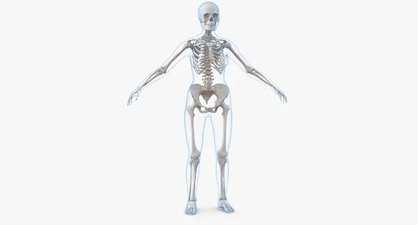
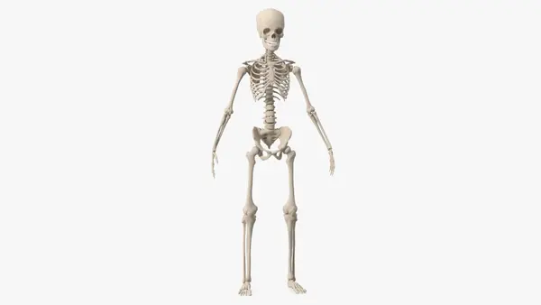
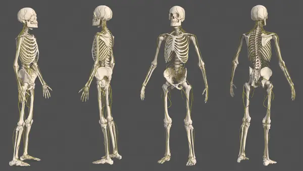
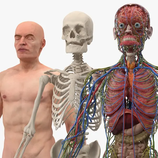
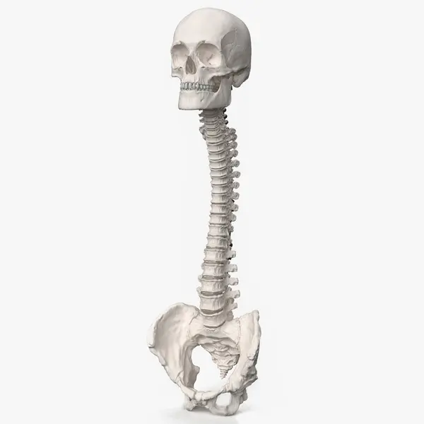
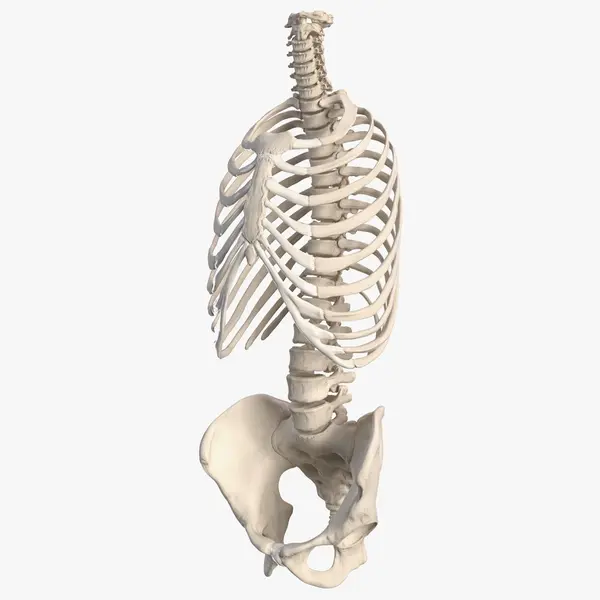
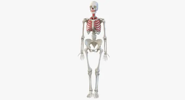
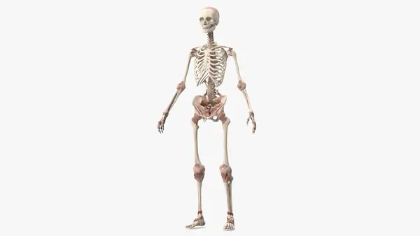
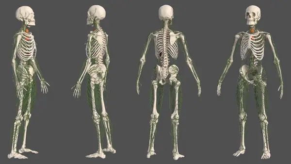
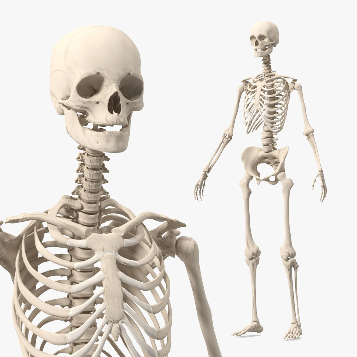
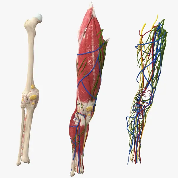
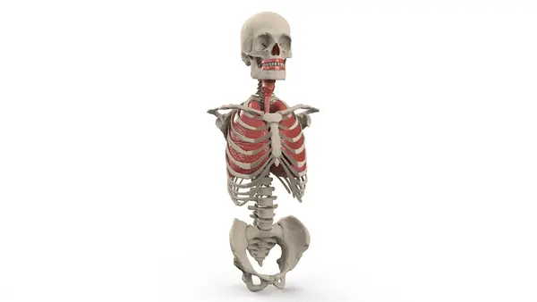
In a skeleton the proportion carries the anatomy. The complete female skeleton makes every bone a separate object, so the ribs can be separated, the pelvis isolated and the vertebrae counted, with 27 maps across 80,733 polygons. The full male skeleton keeps each vertebra, rib and finger joint distinct and carries 75 maps at 8192 x 8192 and 4096 x 4096, an unusually rich set for bone. A skeleton with internal organs shows the ribcage over the lungs and heart and the pelvis around the lower organs. That pairing of layers teaches spatial relationships most clearly.
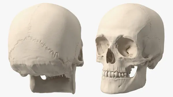
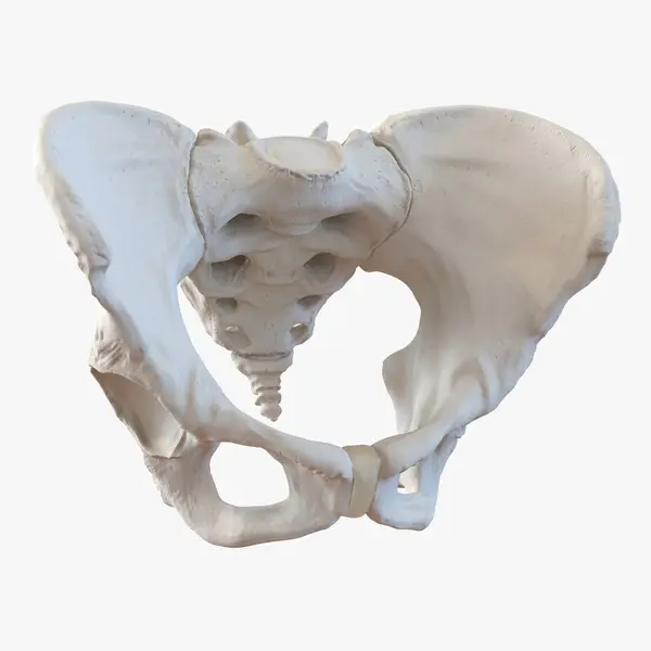
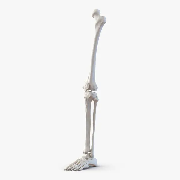
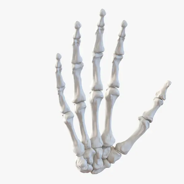
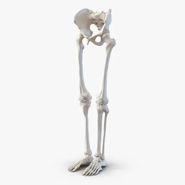
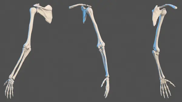
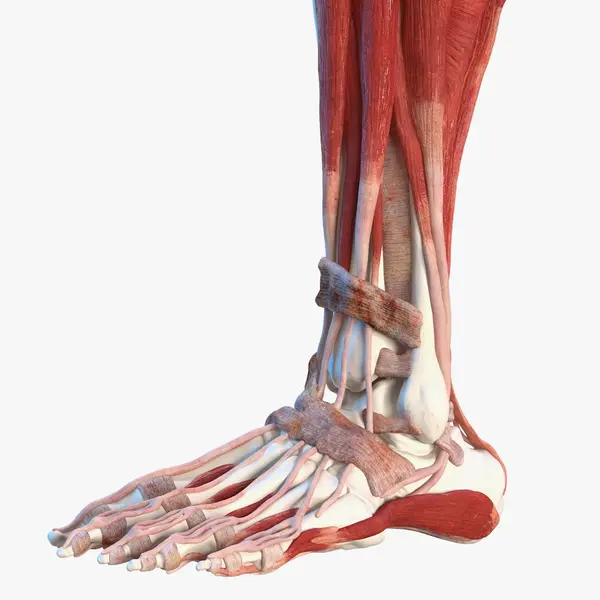
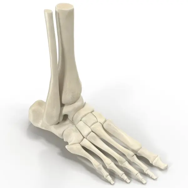
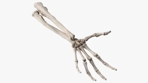
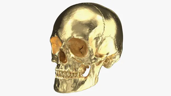
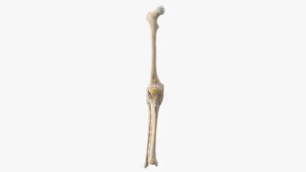
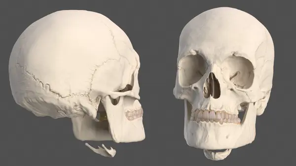
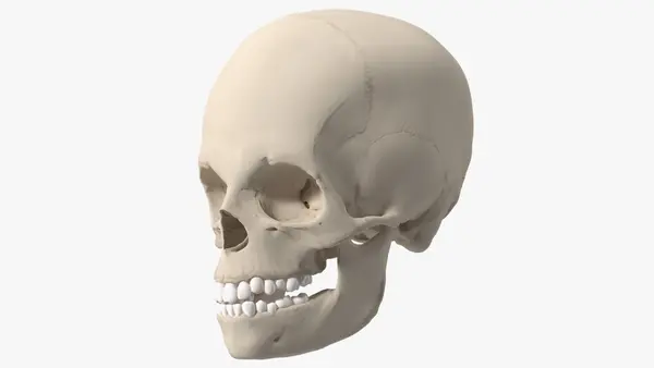
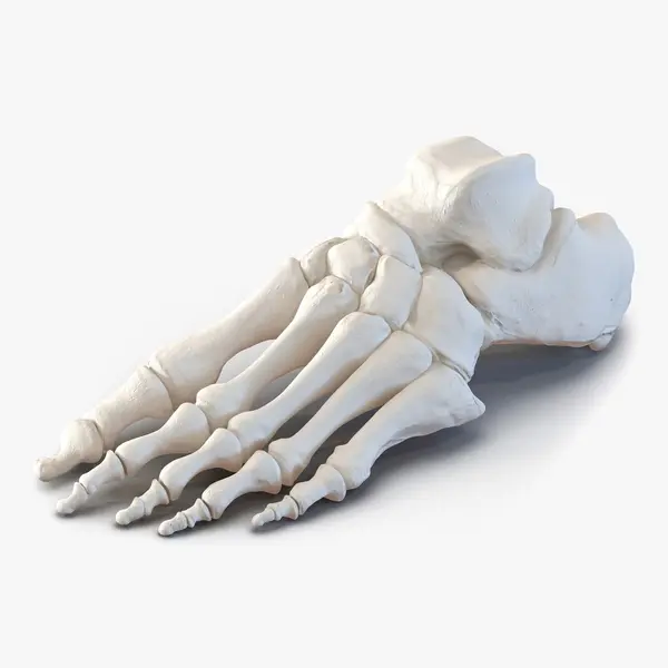
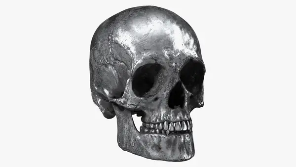
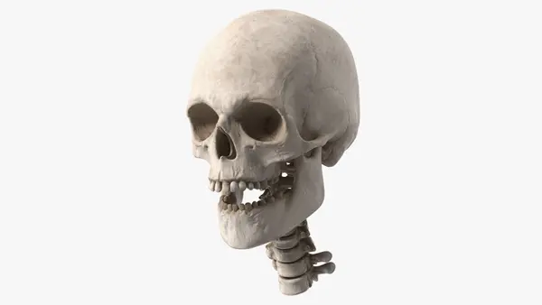
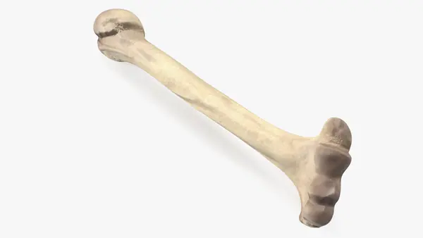
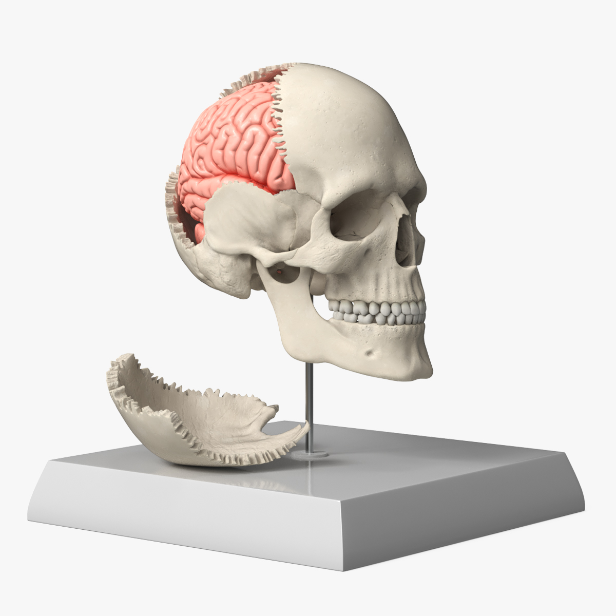
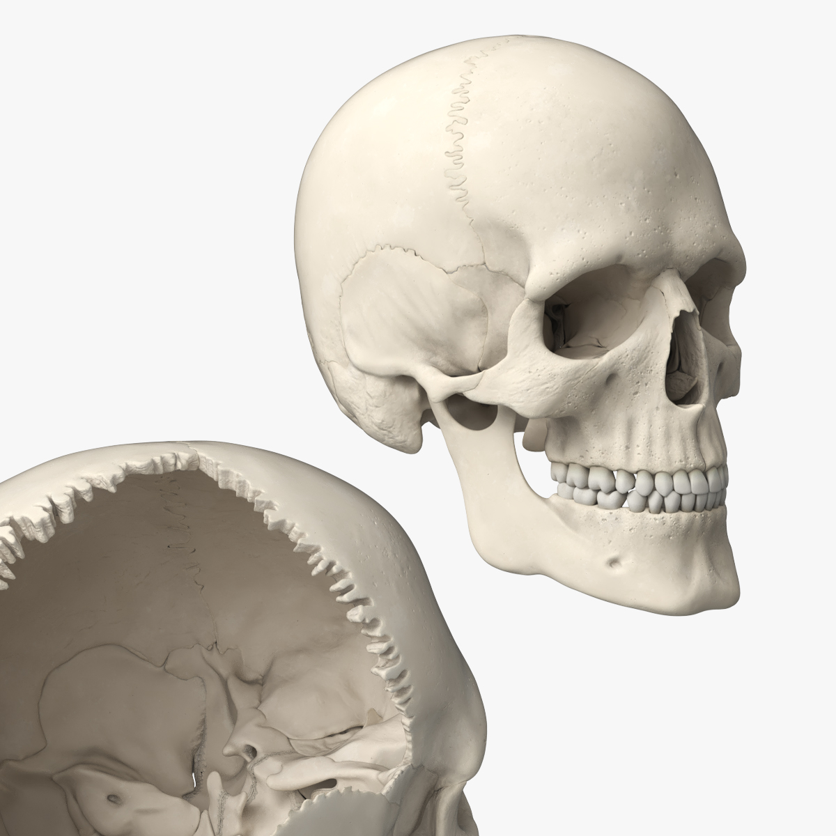
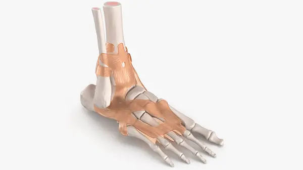
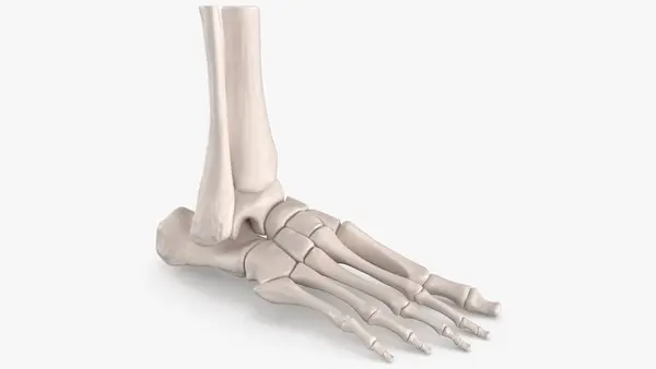
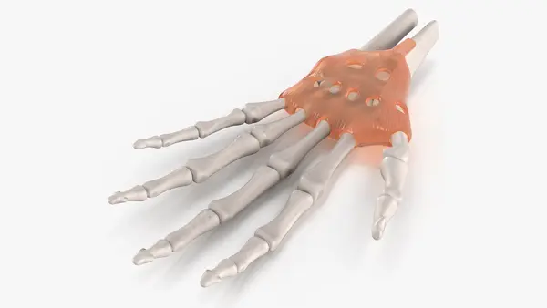
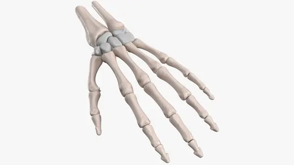
Regional models stay light for web viewers and apps. The hand bones need 3,958 polygons, the leg bones from hip to toes 8,186, and the foot skeleton holds its natural arch in 15,065. A hand skeleton with the flexor retinaculum shows the roof of the carpal tunnel, and the female pelvis spends its 4,960 polygons on the oval inlet and the flared iliac wings. The disassemblable skull lifts off its cap and lower jaw to reveal the inner surface of the vault.
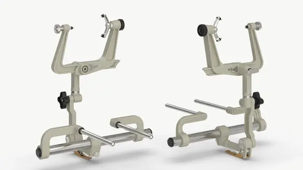
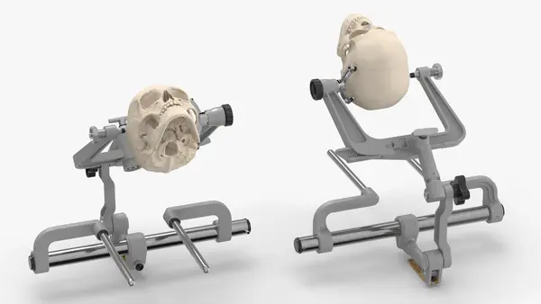
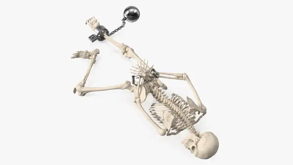
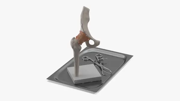
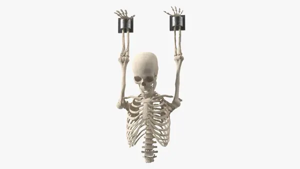
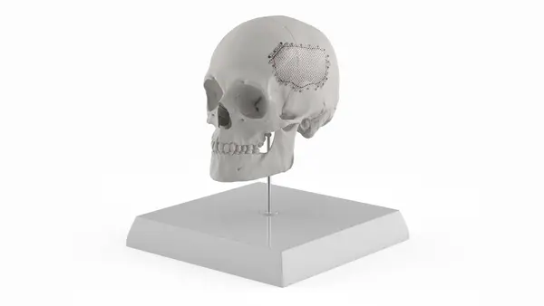
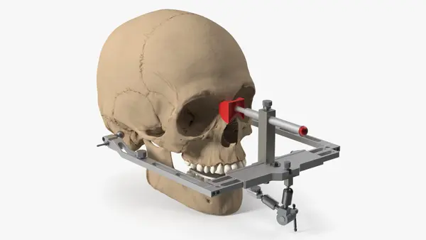
Surgical and story pieces broaden the use. A three-pin skull clamp reaches 7,569,760 polygons, a display skull shows a mesh implant closing a defect, and a shackled skeleton slumped against a wall dresses a dungeon. Anatomy classes, orthopedic and physiotherapy explainers, forensic scenes and patient communication make up most of the work for this Medical subcategory.