Human Anatomy 3D Models
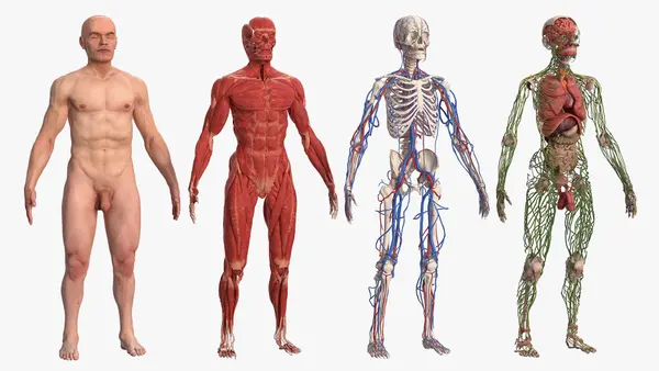
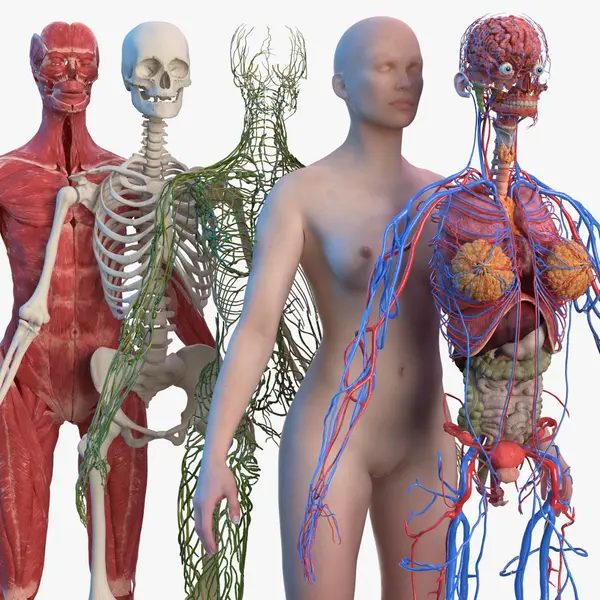
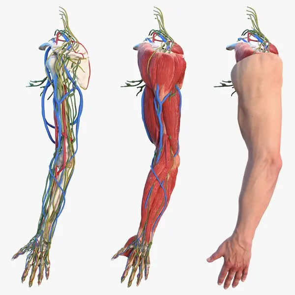
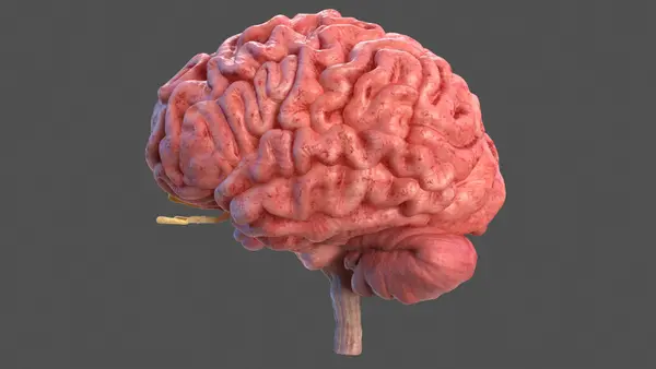
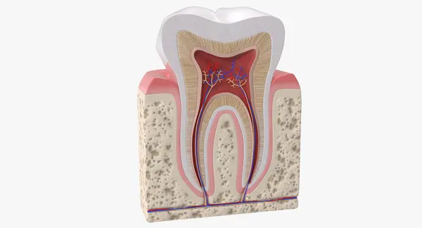
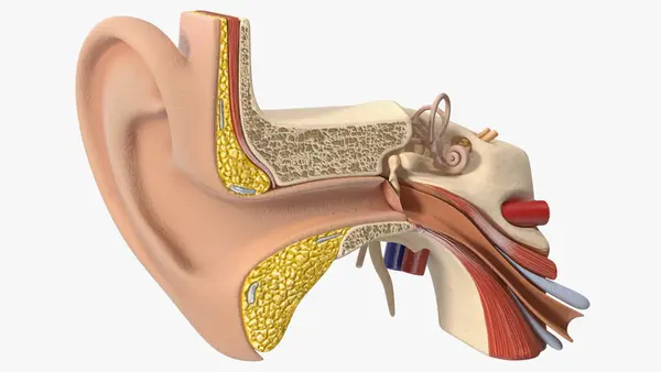
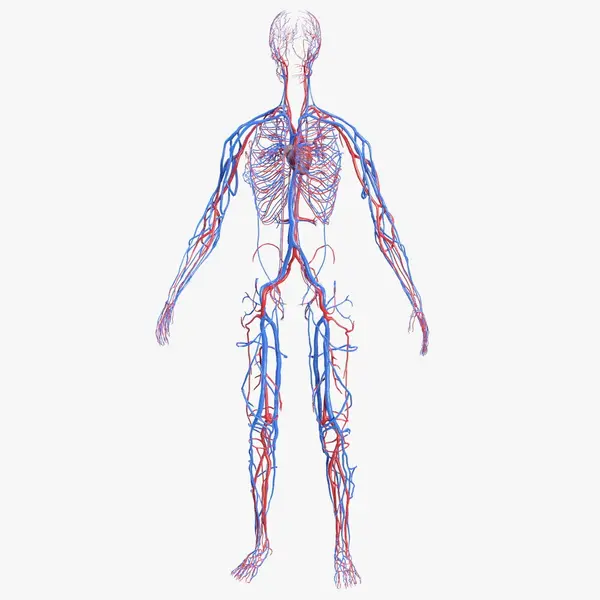
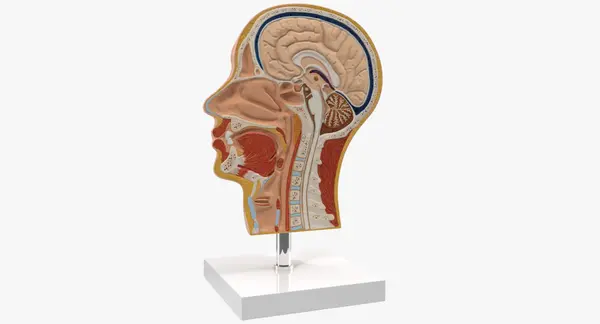
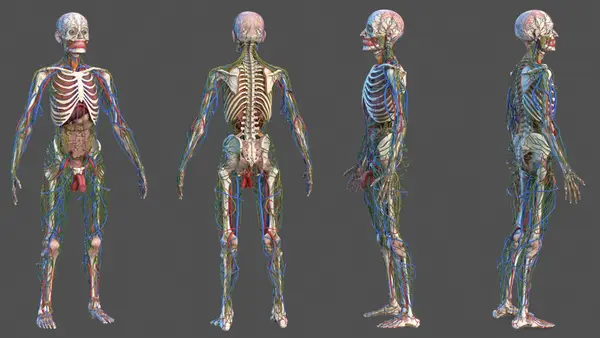
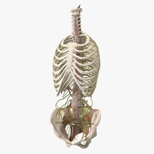
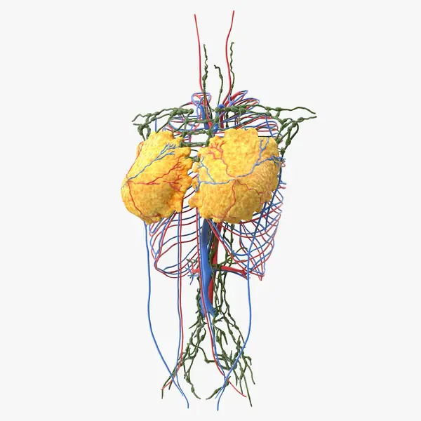
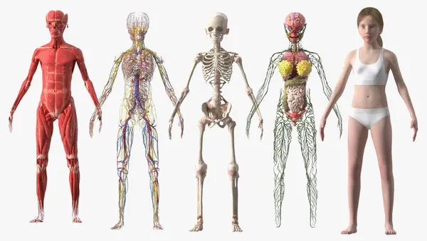
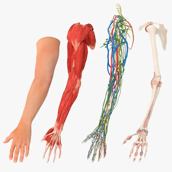
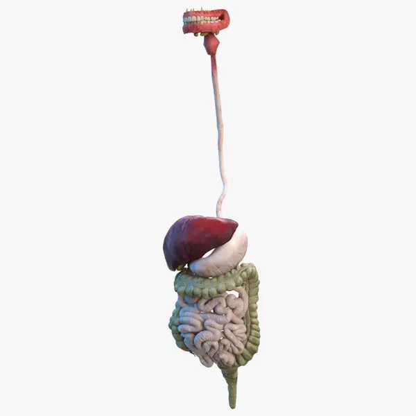
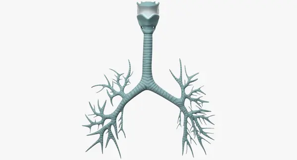
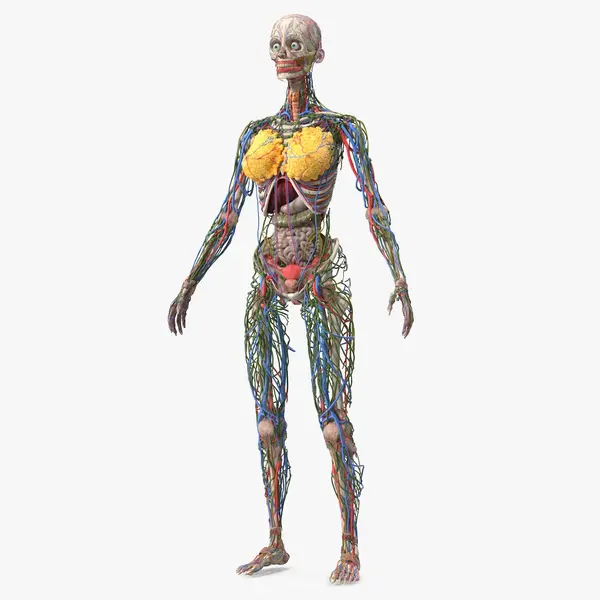
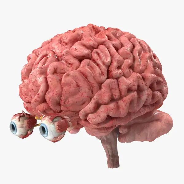
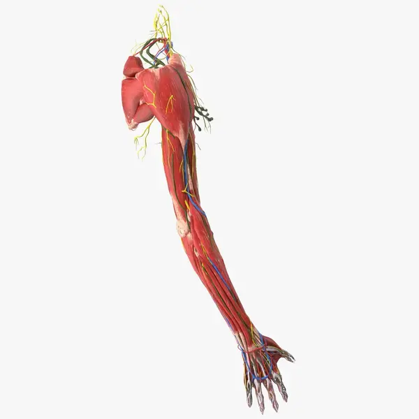
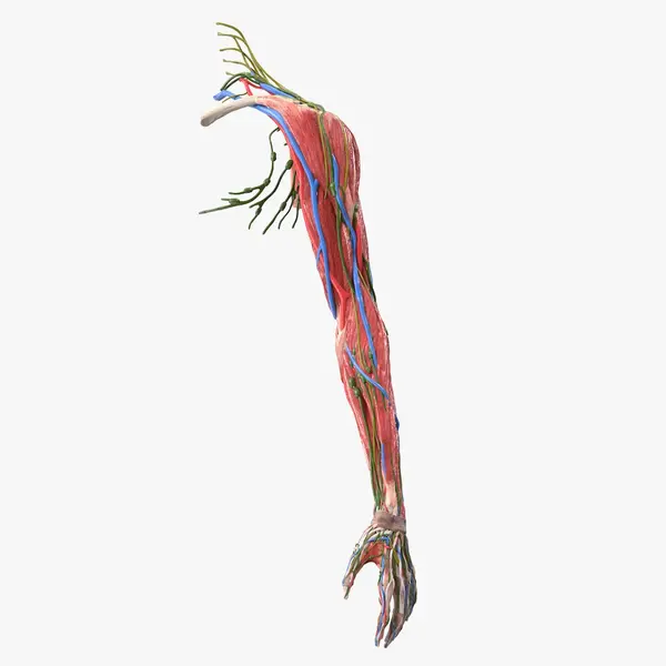
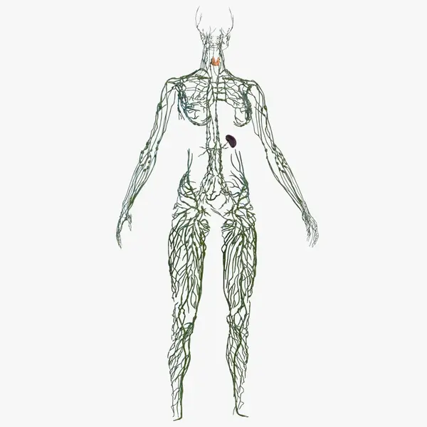
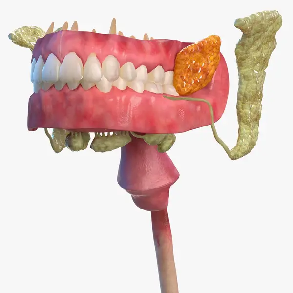
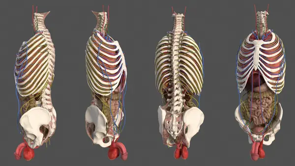
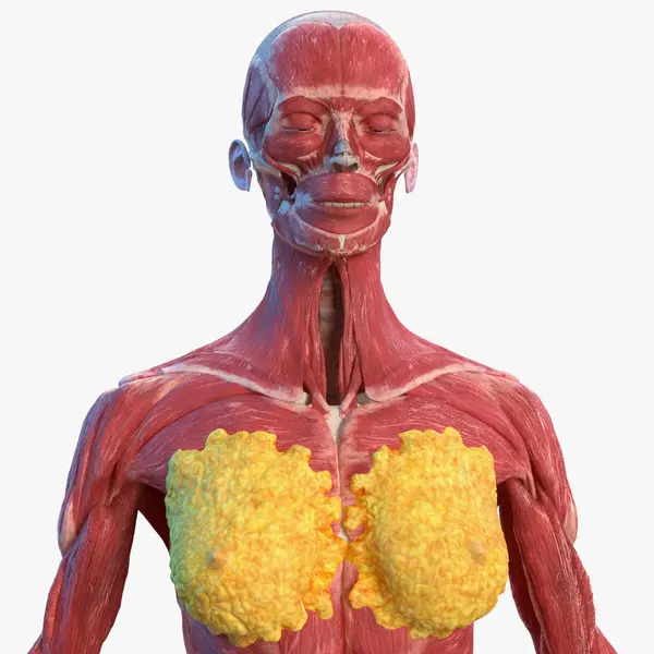
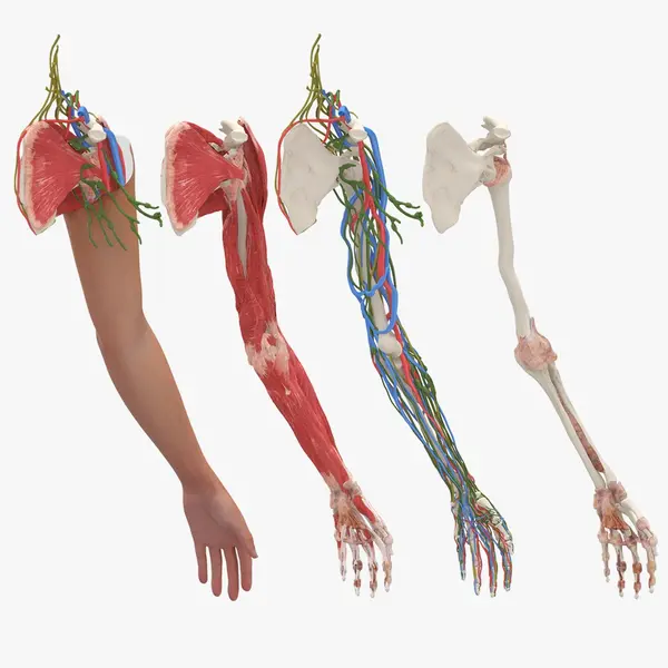
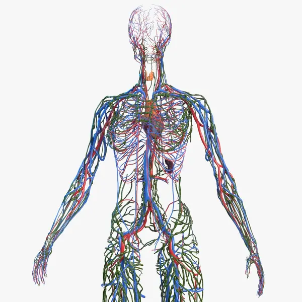
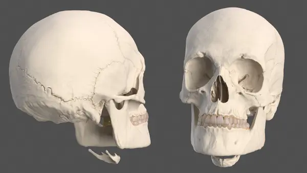
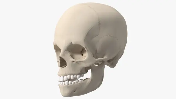
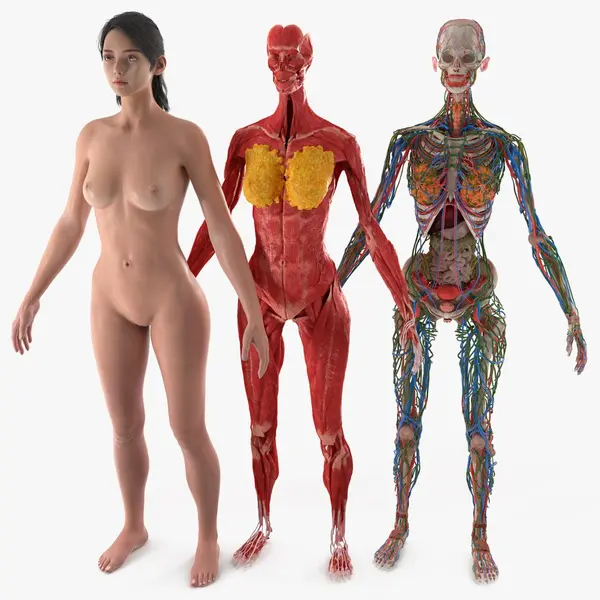
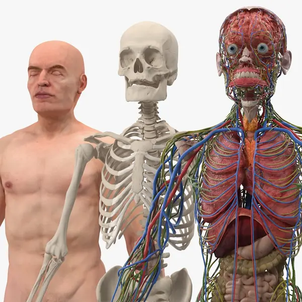
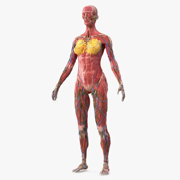
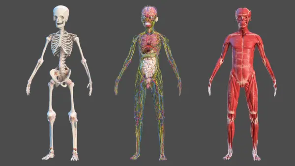
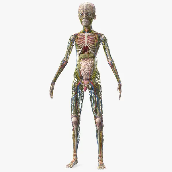
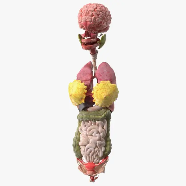
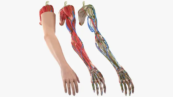
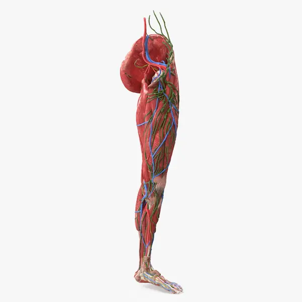
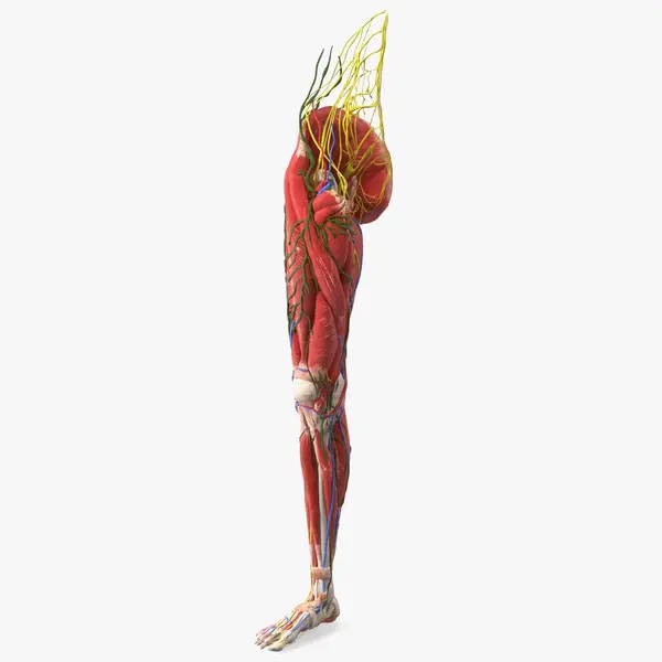
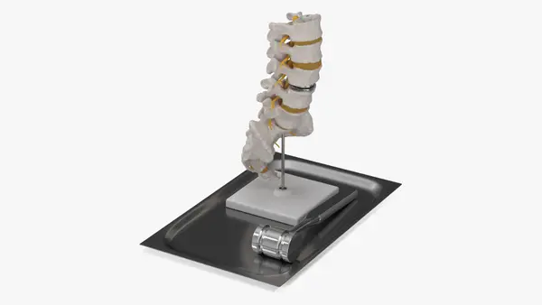
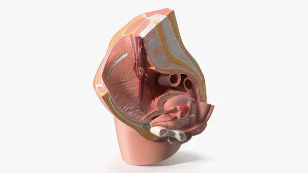
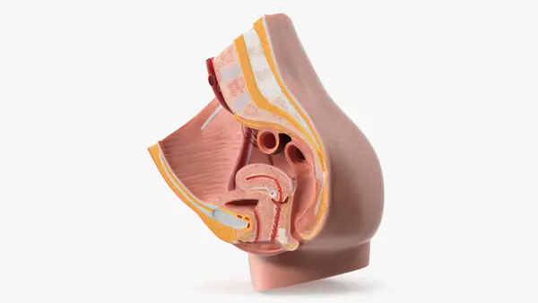
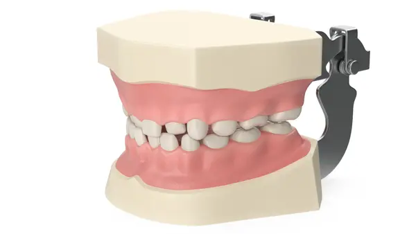
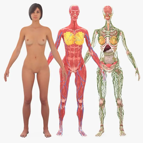
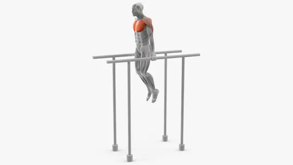
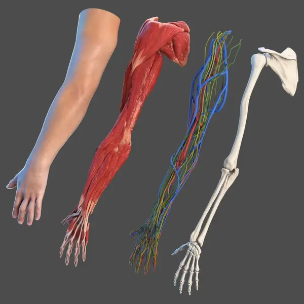
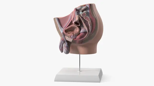
Anatomy models for teaching and medical explanation: full-body figures with skin and organs, heads and limbs built layer by layer, and single organs such as the brain, eye, ear, lungs and urinary tract. Smaller pieces include a nerve cell, knee and shoulder joints, breast cutaways and a skin block on a stand.










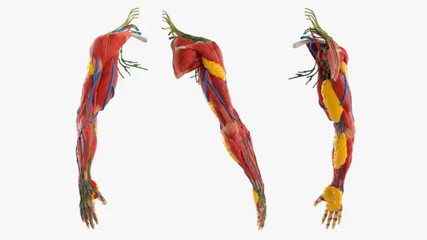



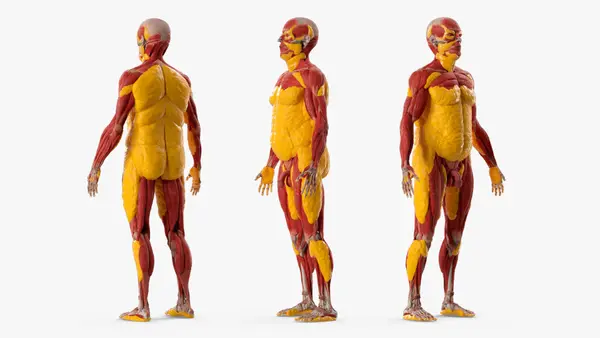
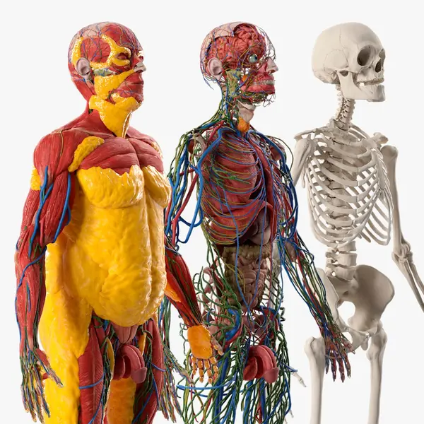
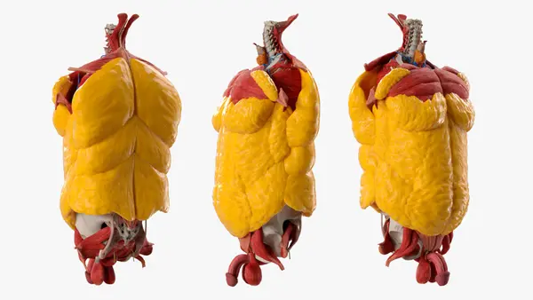
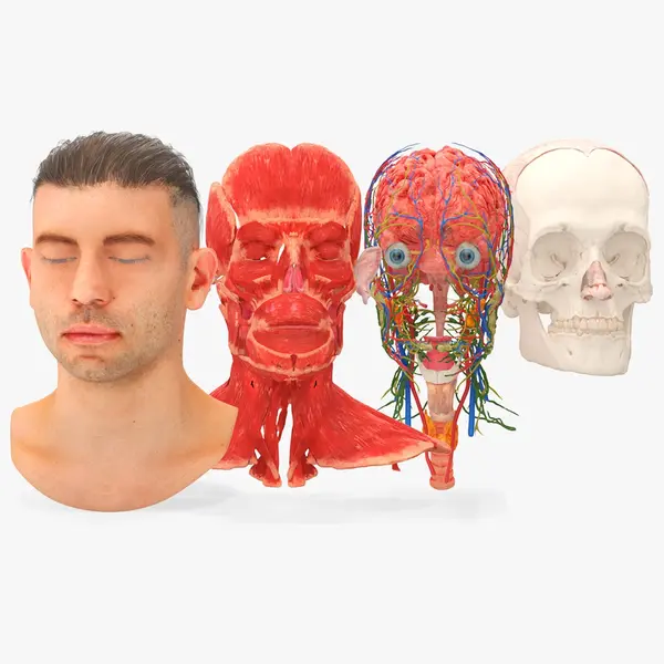


About Human Anatomy 3D Models
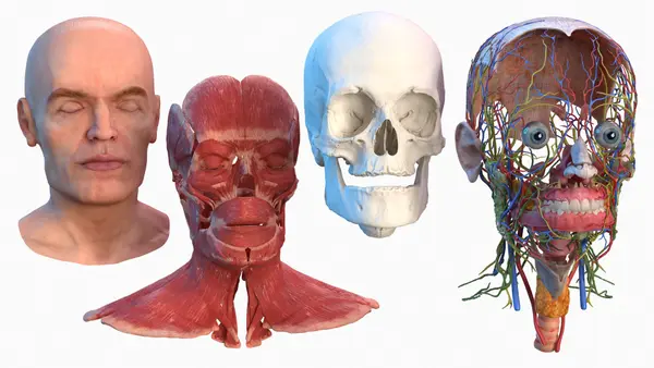
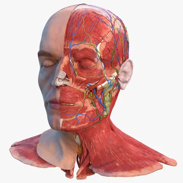
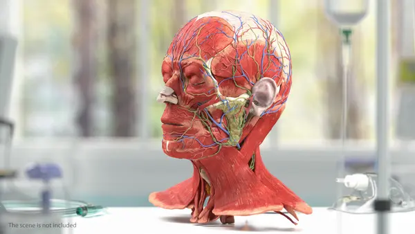
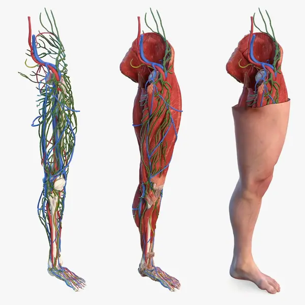
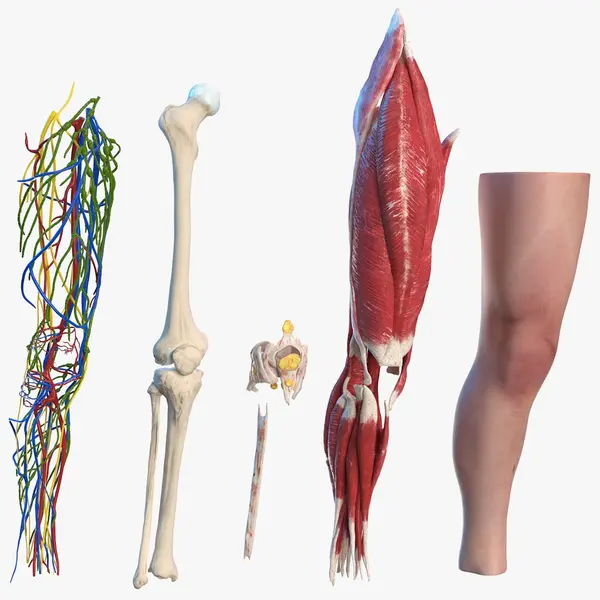
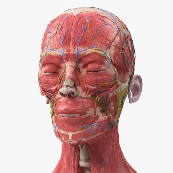
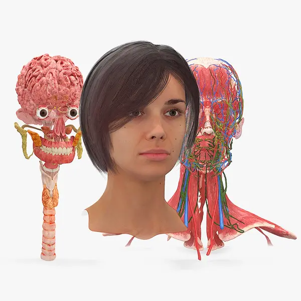
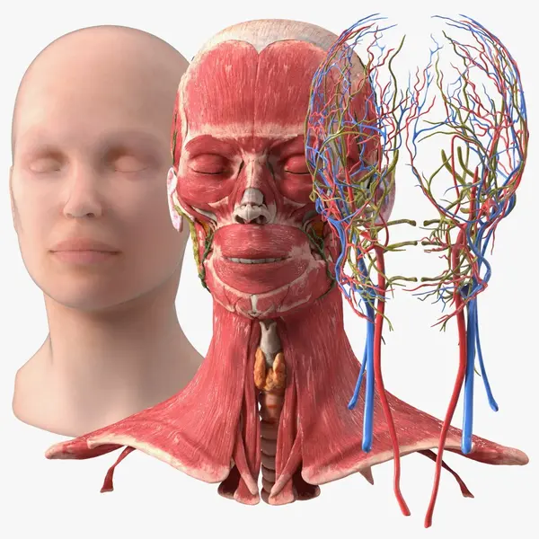
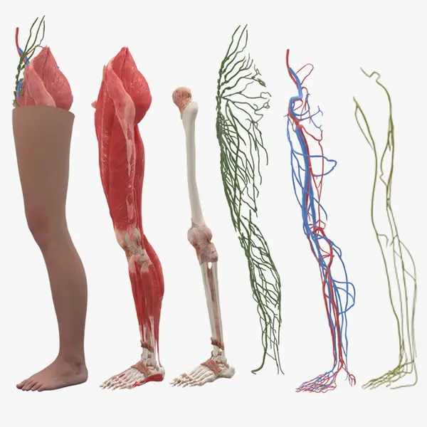
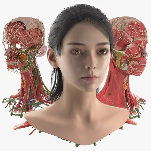
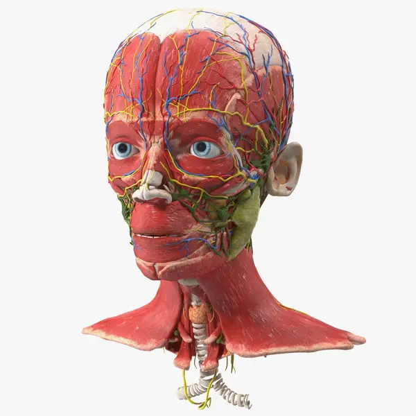
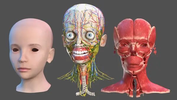
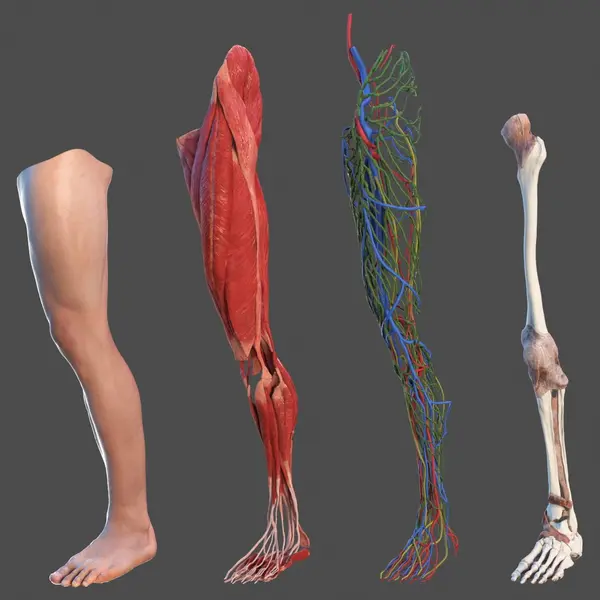
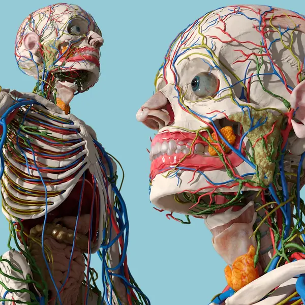
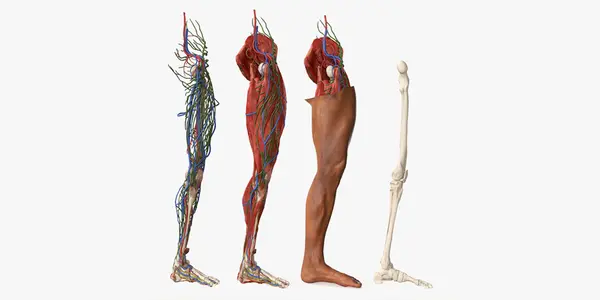
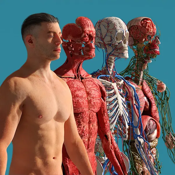
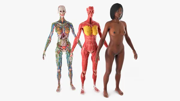
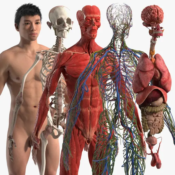
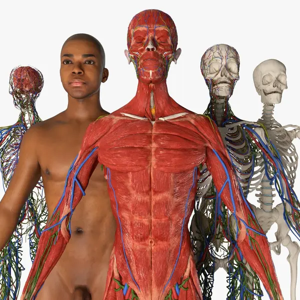
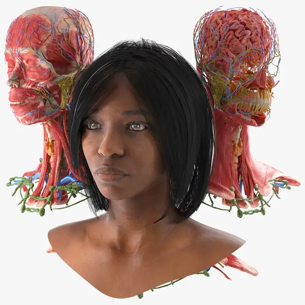
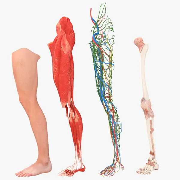
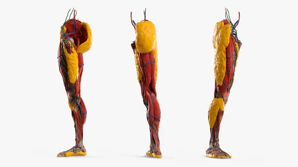
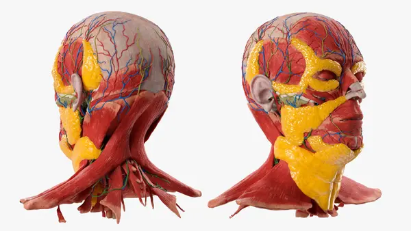
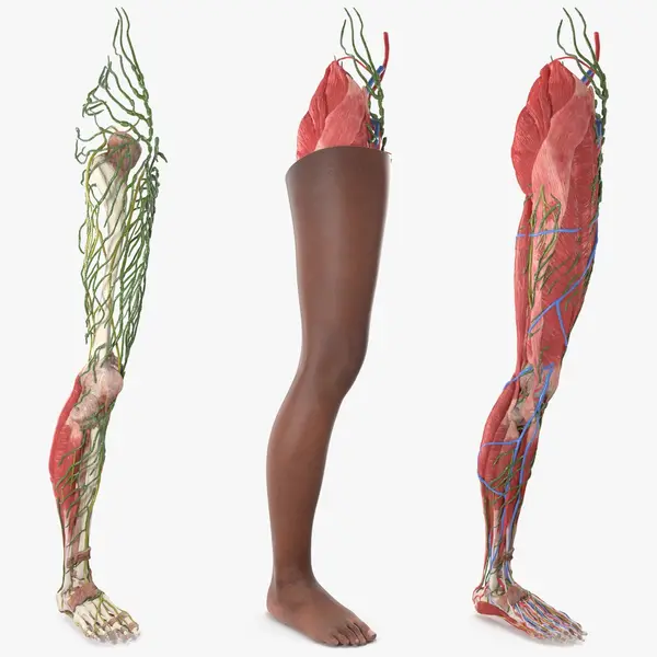
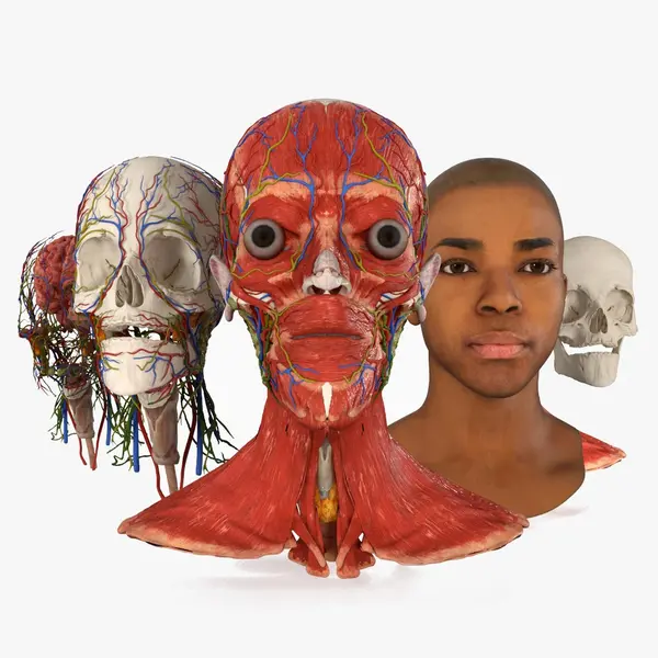
A head with both anatomy and skin is two assets in one: the face gives context, and the muscles, bones and vessels beneath it carry the explanation. Fading between them only works when the skin sits correctly over the structures, and this head uses 312 texture maps to keep each part distinct. The skinless head and neck takes the same approach with 311 maps, so a muscle group, vessel or gland can be isolated and labeled instead of lit as one red mass. The leg with skin answers why the surface looks the way it does: the hollow behind the knee and the line down the shin follow from what sits underneath.
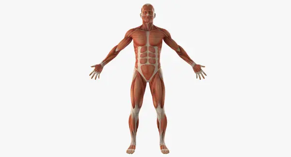
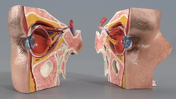
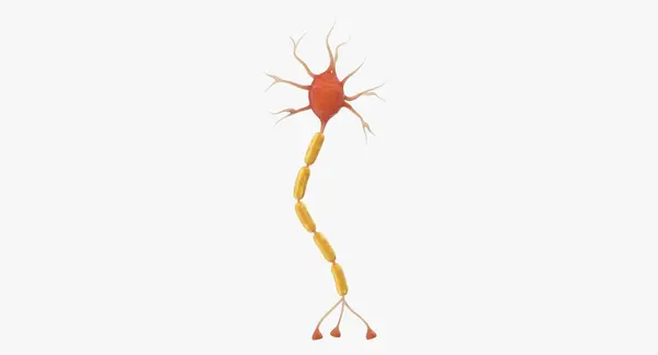
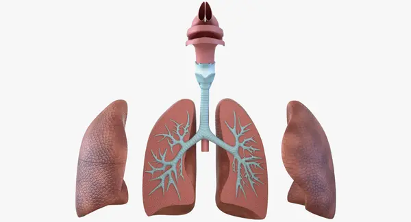
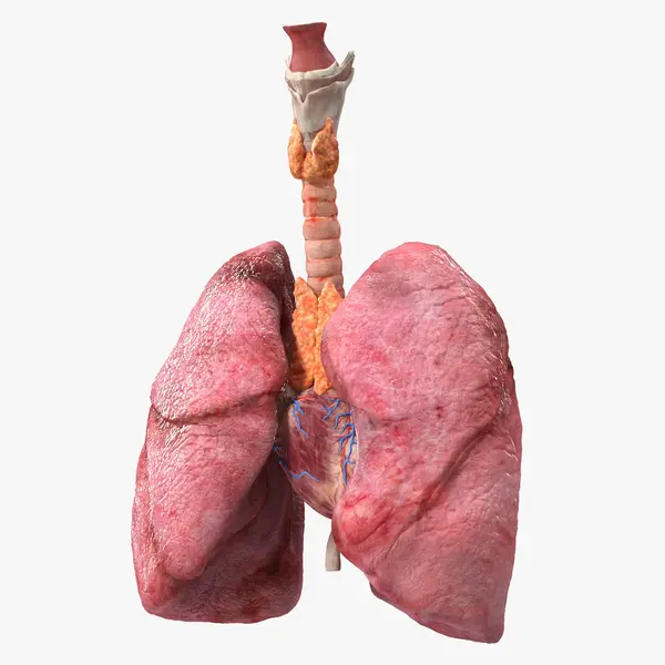
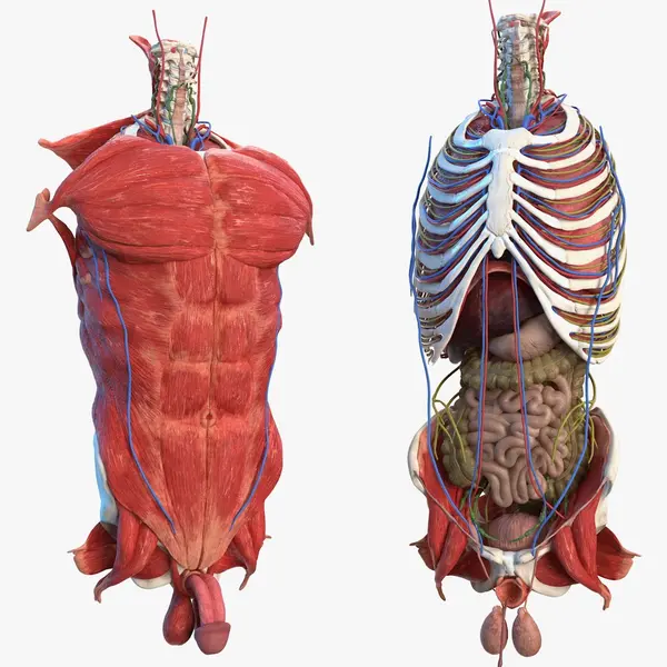
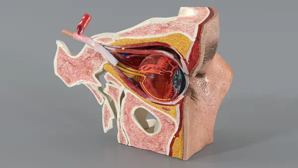
Other models are light by design. The muscular system without a skeleton runs on 7,408 polygons and shows origins and insertions clearly. The lungs use 15,902 polygons and five maps, enough for browser lessons, and they keep the left lung with two lobes rather than three. A nerve cell needs only 2,429 polygons, and a pregnancy breast model is built from 946 for mobile courseware. The sectioned eye shows the lens behind the iris, the vitreous, the retina and the optic nerve.
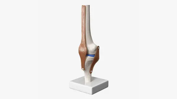
Within the Medical category, choose by layers and weight: skin with anatomy for fades, skinless models for muscle work, single organs for focused lessons. A knee model is prepared for 3D printing, with the bones as solid pieces. Typical users are medical schools, patient education, explainer animation and interactive courseware.